Are you worried because of keratoconus? If so, your first step should be to make an appointment with a trusted optometrist to begin your keratoconus treatment in Madrid. Keratoconus is a condition where the cornea, the eye’s outermost layer and lens, starts to become irregular and bow forward. The cornea plays a crucial role in controlling the quality of light that gets into your eye—and focusing that light onto the retina so that your brain can adequately process visual information—. Any shape change or weakening of this surface can result in significant vision disruption. Specifically, if you are experiencing Keratoconus, you will notice that your vision gets blurrier over time. Most patients with Keratoconus develop the condition in their teens or twenties. And then they will experience a progression of irregular astigmatism over the next decades.
Symptoms of Keratoconus
Keratoconus is not the only eye condition that results in astigmatism. In fact, astigmatism occurs as a result of many irregularities in the cornea’s shape that prevents light from focusing directly onto the retina. As such, the symptoms that Keratoconus brings about—blurry vision, distortion, glare, eye strain, headaches, etc.—are the same symptoms you would likely experience with any other cause for irregular astigmatism. How do you know whether the root of the issue is Keratoconus? Again, your first step should be to see an eye care practitioner and undergo a thorough eye exam.
In Keratoconus, the weakening of the cornea ultimately causes the corneal surface to become thinner and thinner over time. Eventually, the cornea becomes so weak that it can no longer match the eyeball’s spherical shape and starts to bulge outward in a cone-like shape (hence the name “keratoconus”). The more conical the cornea becomes the more severe the symptoms, ranging from nearsightedness to extreme glare around lights and visual distortion—particularly in nighttime or low-light conditions. One symptom patients won’t experience is pain. Though many people diagnosed with Keratoconus assume that the bulging of the cornea will be painful, pain is not a common symptom.
Getting Diagnosed for Keratoconus
There are several methods that an eye care practitioner can use to diagnose Keratoconus beyond asking the patient about symptoms or noticing a conical corneal shape as part of a basic eye exam. One strategy your eye doctor may try is a refractive eye exam. The optometrist uses a retinoscope with a phoropter to test how effectively different lenses are for correcting your vision issues. This test is helpful for diagnosing any refractive error, including myopia (nearsightedness), hyperopia (farsightedness), astigmatism, or presbyopia (age-related farsightedness). Still, it can help diagnose Keratoconus due to the unique way light reflects in this condition.
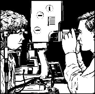
Other tactics include slit-lamp exams, keratometry tests, and corneal mapping (corneal topography). In a slit-lamp exam, your eye doctor will use a beam of light and a microscope to examine your eye, including the shape of your cornea. This method can help spot early thinning of the cornea. In keratometry, an eye doctor directs a circle of light onto your cornea’s surface and uses the light’s reflection to assess your corneal curvature.
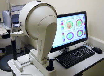
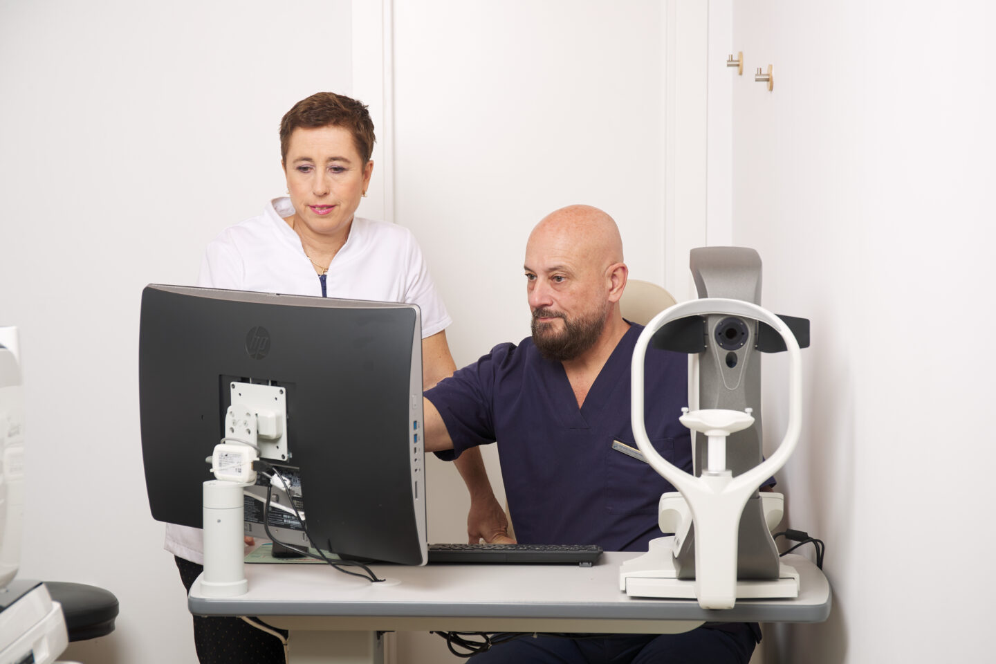
Finally, in corneal mapping or topography, your optometrist will use photographic technology to record images of your cornea and assemble tomographic maps of the corneal surface. This computerized mapping process is terrific for ascertaining corneal thickness, cornea shape irregularities, and early keratoconus signs.
Visit Dr Fernandez-Velazquez Today for Keratoconus Treatment in Madrid
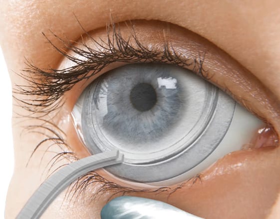
At Centro Fernandez-Velazquez in Madrid, we not only can diagnose keratoconus cases, but we are also experienced in implementing effective keratoconus treatments. Our definitive go-to treatment for Keratoconus is to use scleral lenses. These custom-designed contact lenses rest on the white of the eye (the sclera) and then vault over the corneal surface. This design, which protects the ocular surface by leaving a tear-filled chamber between the eye and the contact lens, helps compensate for irregularities in corneal shape, including anomalies caused by Keratoconus. Our experience has been that scleral lenses are the best option for providing comfort and precise, stable vision for patients with Keratoconus. . You can check our areas of speciality and qualifications here.
Call us today at 915 417 419 to schedule your first in-person consultation. If you need further information, you can contact our office here.

Dr. Fernando Fernandez-Velazquez was born in Madrid, and attended University of Madrid, where he majored in Optics. He then received his Doctor of Optometry degree at New England College of Optometry located in Boston, Massachusetts.
He continued his optometry education and completed a program with emphasis in Contact Lenses.

